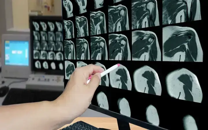
Musculoskeletal Imaging Services
At AMI, our team of highly skilled musculoskeletal radiologists and sports imaging specialists offer screening and diagnostic support with state-of-the-art imaging modalities, cutting-edge technology and the expertise necessary.
The certified subspecialty radiologists at AMI are trained not only to diagnose and treat a wide range of musculoskeletal disorders but also to heal sports injuries. With the latest in Musculoskeletal imaging technology which includes magnetic resonance imaging (MRI), multi-slice CT, and nuclear scintigraphy, we offer the full spectrum of specialized diagnostic services for bones, joints, soft tissue and bone diseases including osteoporosis disorders. Image-guided procedures and spinal injections are used to treat trauma and other orthopedic conditions.
MSK Radiology departments across AMI India & GCC are manned by a trained and dedicated group of image readers. Patients can be scanned on a 3T magnet at AMI outpatient facilities. Selected image-guided aspirations and injections, including steroid injections and arthrography prior to MRI or CT, can be administered. We follow metal artifact reduction protocol on both CT and MRI scans.
We work closely with:
- Orthopedic surgeons
- Physiatrists (physical medicine and rehabilitation specialists)
- Chiropractors
- Rheumatologists

What do we offer at AMI?
How Musculoskeletal Radiology Reporting Can Improve Your Throughput Using Our Services
Quality
Reporting standards followed as per guidelines from the American College of Radiology (ACR) & The Royal College of Radiologists (RCR)
On-Time Reports
Reliable, and accurate reports with less turn-around time. 99% of the emergency reports are delivered in less than 1 hour.
24/7 Compliance
Internationally certified radiologists with Sub-specialty expertise are available 24×7 for 365 days a year.
FAQs
Musculoskeletal imaging is a specialized branch of radiology focused on the comprehensive evaluation of the musculoskeletal system, which includes bones, joints, muscles, and soft tissues. This field employs various imaging modalities such as X-rays, CT scans, MRI, and ultrasound to diagnose and manage a wide range of musculoskeletal conditions and injuries. Musculoskeletal imaging plays a pivotal role in identifying fractures, arthritis, tumors, ligament injuries, and other disorders affecting the bones and joints. It is particularly valuable for assessing sports injuries, degenerative joint diseases, and congenital abnormalities. The detailed anatomical information provided by musculoskeletal imaging aids in treatment planning, guiding orthopedic surgeries, and facilitating the ongoing management of musculoskeletal disorders. Radiologists specializing in musculoskeletal imaging work closely with orthopedic surgeons, rheumatologists, and other healthcare professionals to ensure accurate diagnoses and optimal patient care, contributing significantly to the overall field of musculoskeletal medicine.
Magnetic Resonance Imaging (MRI) is widely regarded as the best imaging modality for the musculoskeletal system. This non-invasive technique provides detailed, high-resolution images of soft tissues, bones, and joints, making it particularly effective in diagnosing a diverse range of musculoskeletal conditions. MRI offers superior contrast resolution, allowing for the precise visualization of ligaments, tendons, muscles, and articular surfaces. It is highly sensitive in detecting abnormalities such as fractures, joint disorders, tumors, and inflammatory conditions. Additionally, MRI does not involve ionizing radiation, which is advantageous for repeated imaging studies or when assessing conditions in paediatric patients. The versatility of MRI in showcasing both structural and functional aspects of the musculoskeletal system, along with its ability to capture multiplanar images, makes it an indispensable tool for orthopedic and rheumatologic diagnoses, treatment planning, and monitoring therapeutic outcomes.
Ultrasound imaging plays a significant role in the diagnosis of musculoskeletal injuries, offering a real-time and dynamic assessment of soft tissues, muscles, tendons, ligaments, and joints. This non-invasive imaging modality utilizes high-frequency sound waves to create detailed images, providing valuable insights into the anatomy and function of the musculoskeletal system. Ultrasound is particularly advantageous in the evaluation of injuries, as it allows for dynamic examinations, enabling clinicians to assess structures in motion, such as tendons during joint movement. It is commonly used to diagnose conditions like tendonitis, muscle tears, ligament injuries, and fluid accumulation within joints. The ability to perform real-time imaging during procedures like needle aspirations or injections enhances its utility for interventions. Ultrasound is portable, cost-effective, and does not involve ionizing radiation, making it suitable for point-of-care assessments and repeated examinations. While it may not provide the same depth of penetration as other modalities like MRI, ultrasound remains a valuable tool in the initial assessment and follow-up of musculoskeletal injuries, contributing to a comprehensive and dynamic approach in the diagnosis and management of such conditions.
Musculoskeletal imaging employs several modalities to comprehensively assess and diagnose conditions affecting the bones, joints, muscles, and soft tissues. X-rays are commonly used as an initial imaging tool, offering a quick and cost-effective assessment of bone structure and alignment. Computed Tomography (CT) scans provide detailed cross-sectional images and are particularly useful for evaluating complex fractures and bony abnormalities. Magnetic Resonance Imaging (MRI) is highly effective in capturing detailed images of soft tissues, making it the modality of choice for assessing ligaments, tendons, and joint structures. Ultrasound, utilizing sound waves, offers real-time imaging, making it valuable for dynamic assessments of muscles, tendons, and joints, especially during movement. Nuclear medicine techniques, such as bone scintigraphy, help identify areas of increased or decreased bone metabolism, aiding in the diagnosis of conditions like fractures and tumors. Each modality has its strengths and is chosen based on the specific clinical scenario, contributing to a comprehensive approach in musculoskeletal imaging for accurate diagnoses and effective treatment planning.

AMI Expertise - When You Need It, Where You Need It.
Partner With Us

