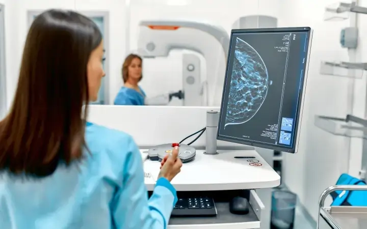
Breast Imaging And Mammography Radiology
At AMI, our team of highly specialized radiologists delivers screening and diagnostic support with state-of-the-art imaging modalities and advanced technology. The radiology subspecialty is devoted to diagnostic imaging including mammography, breast ultrasound, breast MRI for screening and early diagnosis of breast pathologies thereby enhancing medical and surgical management.
The Breast Imaging and Mammogram team at AMI is the most subspecialized group of breast imaging radiologists in India and the GCC region with expertise in breast ultrasound, MRI, and biopsy techniques.
Breast imaging is often considered to be limited to screening for breast cancer. However, this subspecialty includes diagnosis of all breast conditions using mammograms, breast ultrasounds, image-guided aspirations and biopsies, magnetic resonance imaging (MRI), ductography, and molecular imaging.
95% of breast diseases occur in women and are rarely seen in men. While early detection is key to the treatment and cure of breast diseases, mammography radiology plays a vital role in its diagnosis. Our team of highly skilled and experienced radiologists uses advanced, high-resolution imaging equipment to diagnose and treat diseases — helping many women achieve better, long-term health outcomes.
Our breast imaging radiologists work closely with:
- Primary care physicians
- Obstetricians and gynecologists
- Breast surgeons
- Medical oncologists
- Radiation oncologists

What do we offer at AMI?
How Breast Imaging & Mammography Reporting Can Improve Your Throughput Using Our Services
Quality
Reporting standards followed as per guidelines from the American College of Radiology (ACR) & The Royal College of Radiologists (RCR)
On-Time Reports
Reliable, and accurate reports with less turn-around time. 99% of the emergency reports are delivered in less than 1 hour.
24/7 Compliance
Internationally certified radiologists with Sub-specialty expertise are available 24×7 for 365 days a year.
FAQs
Mammogram and breast imaging are both techniques used to visualize the breast tissue, but they differ in their specific modalities and purposes. A mammogram specifically refers to a type of breast imaging that utilizes low-dose X-rays to create detailed images of the breast tissue. Mammograms are primarily used for breast cancer screening, detecting abnormalities such as tumors or calcifications, and monitoring changes in breast health over time. On the other hand, breast imaging is a broader term that encompasses various imaging techniques used to assess the breasts, including mammography, ultrasound, magnetic resonance imaging (MRI), and molecular breast imaging (MBI).
Breast imaging may involve additional modalities beyond mammography, such as ultrasound or MRI, to further evaluate breast abnormalities detected on mammograms or to assess specific clinical concerns. While mammography remains the primary screening tool for breast cancer, breast imaging encompasses a range of techniques tailored to individual patient needs and clinical scenarios, providing comprehensive assessment and management of breast health.
Breast imaging radiology refers to the specialized field within radiology focused on the imaging and evaluation of the breast tissue. Radiologists trained in breast imaging use various imaging modalities to assess breast health, detect abnormalities, and aid in the diagnosis and management of breast diseases, particularly breast cancer. Common imaging techniques used in breast imaging radiology include mammography, ultrasound, magnetic resonance imaging (MRI), and molecular breast imaging (MBI). Mammography, utilizing low-dose X-rays, is the primary screening tool for breast cancer and is highly effective in detecting early-stage tumors and calcifications. Ultrasound is valuable for further evaluating abnormalities detected on mammograms and assessing breast masses, while MRI provides detailed images of breast tissue and is often used in high-risk patients or for staging purposes. Molecular breast imaging involves the injection of a radioactive tracer to detect breast abnormalities. Breast imaging radiologists play a critical role in interpreting imaging studies, providing accurate diagnoses, guiding treatment decisions, and monitoring treatment responses, contributing significantly to the comprehensive care of patients with breast diseases.
Another approach to determining the best type of breast imaging involves considering the individual patient's characteristics and specific clinical needs. For instance, in women with dense breast tissue, which can make mammography less sensitive, supplemental screening with breast ultrasound or MRI may be recommended to improve cancer detection rates. Additionally, for women at high risk of breast cancer due to factors such as genetic mutations or strong family history, MRI is often preferred due to its high sensitivity for detecting tumors. In cases where there is a need for further evaluation of suspicious findings or for guiding biopsy procedures, technologies such as ultrasound or MRI may offer advantages over mammography. Ultimately, the best type of breast imaging varies depending on the patient's risk factors, breast density, and the clinical question at hand, highlighting the importance of personalized decision-making in breast cancer screening and diagnosis. Consulting with a breast imaging specialist can help determine the most appropriate imaging approach for each individual patient.
Mammography is widely regarded as the gold standard for breast cancer screening due to its proven effectiveness in detecting early-stage tumors and calcifications, often before they can be felt. It is a low-cost, non-invasive imaging technique that utilizes low-dose X-rays to produce detailed images of the breast tissue. Mammography has been extensively studied and has demonstrated a significant reduction in breast cancer mortality through early detection and intervention. Additionally, mammography is widely accessible, making it feasible for large-scale screening programs aimed at detecting breast cancer in asymptomatic women. While mammography may have limitations, such as reduced sensitivity in women with dense breast tissue, it remains the most widely utilized and recommended screening tool for breast cancer due to its ability to detect tumors at an early stage when treatment is most effective. Moreover, advancements in technology, such as digital mammography and 3D mammography (tomosynthesis), have further improved the sensitivity and accuracy of mammographic screening, reinforcing its status as the cornerstone of breast cancer detection and prevention efforts.
One of the latest trends in breast imaging radiography is the increasing utilization of abbreviated or rapid breast MRI protocols for breast cancer screening in certain patient populations. These protocols aim to streamline the MRI process, making it faster and more cost-effective while maintaining high sensitivity for detecting breast cancer. Abbreviated breast MRI protocols typically involve shorter scan times and fewer sequences compared to traditional full-length breast MRI exams, making them more accessible and feasible for broader implementation in clinical practice. Studies have shown that abbreviated breast MRI protocols can detect additional cancers in women with dense breast tissue or those at high risk of breast cancer, providing valuable supplemental screening beyond mammography. This trend reflects a growing recognition of the importance of personalized screening approaches tailored to individual patient risk factors and preferences, ultimately improving breast cancer detection and patient outcomes.
Tomography, mammography, and ultrasound imaging are all modalities used for breast cancer detection, each with its own distinct characteristics and advantages. Tomography, particularly digital breast tomosynthesis (DBT), is a type of X-ray imaging that captures multiple images of the breast from different angles, allowing for a three-dimensional reconstruction of breast tissue. DBT provides improved visualization of breast structures and has been shown to reduce false positives compared to traditional 2D mammography. Mammography, on the other hand, involves low-dose X-rays to create two-dimensional images of the breast tissue. Mammography is the primary screening tool for breast cancer and is highly effective in detecting calcifications and early-stage tumors. Ultrasound imaging utilizes sound waves to produce real-time images of the breast tissue and is often used as a supplemental imaging modality to further evaluate suspicious findings detected on mammography or to assess breast masses in younger women or those with dense breast tissue. While each modality has its own strengths and limitations, a multimodal approach combining mammography with tomosynthesis and ultrasound can enhance breast cancer detection and improve diagnostic accuracy.

AMI Expertise - When You Need It, Where You Need It.
Partner With Us

